
16 Superior Vena Cava Syndrome (SVCS)
Patient simulation & Special Considerations: SVCS
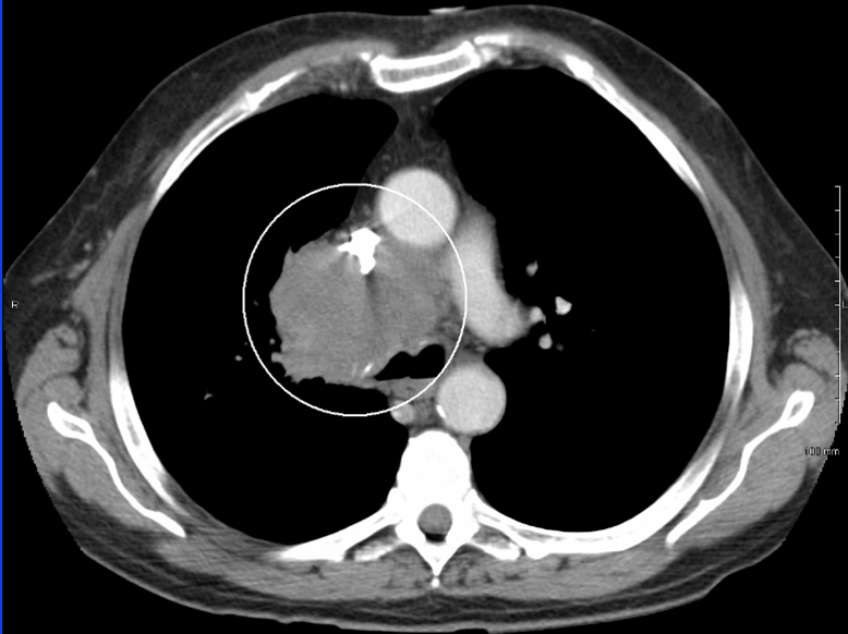
Superior Vena Cava Syndrome is considered a medical emergency; patients must start treatment immediately and receive 2-3 daily fractions. Patients with a SVC obstruction often present with shortness of breath and dyspnea, facial swelling, distension of the veins of the neck and thorax, chest pain, cough, dysphagia, and cyanosis of the upper body. These side effects result from a tumor compressing the thin-walled superior vena cava in the upper chest.
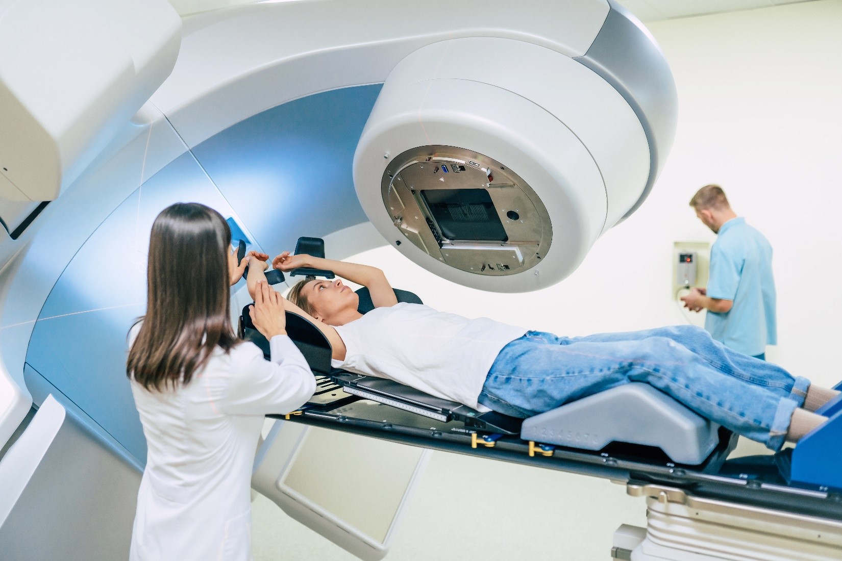
Positioning patients with SVCS is complex because of the respiratory compromise and subsequent orthopnea. Most patients with this condition will require elevation of their upper body using an incline board or custom vacloc or cushion to elevate their head and upper chest. The patient is positioned as close to supine as possible for clinical setups. Some patients may benefit from their doctor prescribing oxygen during their treatment; their oxygen saturation should be monitored throughout simulation and treatment.
Treatment Volume Localization: SVCS
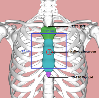
Clinical setups for SVCS start with a 12×12 cm field size. First, palpate the suprasternal notch at the T2-T3 vertebral level and the xiphoid process at the T9-T10 vertebral level and center halfway between. Place the isocenter mid-sternum over the hilum/carina. To find the treatment depth, measure the patient’s thickness at isocenter and divide it by 2; set the SSD. For example. If the patient’s thickness is 20 cm / 2 = 10 cm. The anterior SSD is 90 cm.
The treatment field should include the primary tumor and a 2-3 cm margin. Mediastinal, hilar, and supraclavicular areas are also included. The doctor approves the treatment portal images for every field before treatment. Therapists should document the patients setup with photos and the table and field parameters. A monitor unit calculation should utilize a second check by a physicist or physician.
Treatment Techniques: SVCS
External beam radiation therapy is the treatment of choice for SVCS. The initial 2-3 treatments consist of AP/PA beams with doses of 3-4 Gy/fx given daily until symptoms have improved or a computer-based plan is generated. After the initial treatments, smaller fractions of 1.8-2.0 Gy are delivered daily. The total dose delivered, and future treatment techniques depend on the histology of the tumor. For example, if the SVC is related to a lymphoma, treatment fields may remain the same. If the pathology is lung cancer, an IMRT or VMAT plan may be implemented.
Media Attributions
- SVCS CT image © James Heilman, MD is licensed under a CC BY-SA (Attribution ShareAlike) license
- SVCS positioning © Maksym Povozniuk is licensed under a All Rights Reserved license
- SVCS iso location © Was a bee adapted by Jared Stiles is licensed under a CC BY-SA (Attribution ShareAlike) license
difficulty breathing
difficulty swallowing
Shortness of breath when lying down flat and improves when sitting or standing up
